
CASE OF USELESS XRAY IN SEPSIS...POCUS COMES TO RESCUE
Dr Himanshu Gul Mirani, Specialty Doctor, A&E, QMC
Nottingham University Hospitals, NHS Trust
Case Presentation:
A 70 year old female with h/o CCF was sent to ED, QMC by heart failure district nurse with suspected acute on chronic heart failure and fever. On examination her saturation was 94% on 2L O2 from nasal cannula and
chronic bilateral pedal edema with normal JVP. She had bibasal crackles.
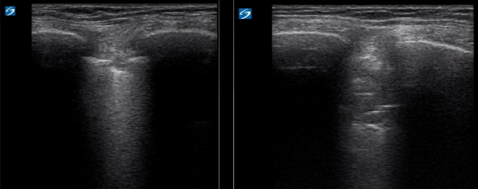
Right chest subpleural changes
Right chest air bronchograms on POCUS
Management and Outcome:
CXR showed left pleural effusion and overlying atelectasis/consolidation as seen in the CXR a month ago.
POCUS scan did not show B line burden suggestive of acute pulmonary edema, instead dynamic air-
bronchograms were seen on the right chest which are highly suggestive of pneumonia. In under 30 mins post registration she had antibiotics tailored to source. Later in her blood tests revealed WCC 19.7 x10^9/L, neutrophils of 17 x10^9/L and CRP of 238.
Key Learnings and Points:
Ultrasound has been shown to be superior to CXR in several studies to identify pneumonia. Ultrasonographic features of pneumonia are dynamic air-bronchograms, focal B lines, sub-pleural consolidation, effusion & hepatization of the lung.
These features when used in the right clinical context, serve a
powerful data point in management of septic patients, thus limiting
broad spectrum antibiotic use; helping in antibiotic stewardship and
reducing drug resistance.